1 · Iron Causes Aging
Aging is a process which causes our bodies to accumulate damage beyond our natural ability to repair it. When our bodies are damaged, they don't function as well, and so aging means an increase in, or a greater tendency to, illness and disease.
Older people have much greater rates of almost every illness than do younger people.
But what causes aging? Scientists have come up with many theories as to how our bodies lose the ability to repair themselves and allow damage to build up. Rather than list all of them, let's look at calorie restriction, since this provides important clues as to how aging works.
Calorie restriction decreases iron and slows aging
Calorie restriction is the most potent, robust intervention for extending the lifespan of laboratory animals; calorie restriction retards aging. By discovering how calorie restriction works, we can gain insight into the aging process.
In calorie restriction, animals are fed from 10% to 50% fewer calories than they would normally eat, or would like to eat, and they live up to 50% longer than normally fed animals. This phenomenon has been tested and proven in a wide range of animals, from microscopic worms (C. elegans), to rodents, to monkeys.
Human data using calorie restriction is somewhat lacking, since humans live much longer than most lab animals, and not many people have been restricting their food long enough to see results; but so far, a number of studies indicate that people who restrict their food intake have much better health.
Much research on calorie restriction has focused on its effect on growth hormone, which it decreases. In addition, calorie restriction decreases inflammation and oxidative damage, improves cell function, and increases autophagy (the cellular self-cleaning and recycling process). All of these processes are strongly associated with aging, so retarding them slows aging.
Calorie restriction also results in a much lower level of iron in aged animals. This may be one of the most important ways that it slows aging.
Lab rats that are food restricted by 40% accumulate far lower levels of iron in their bodies as they age.8viii The rats also have much lower levels of aging damage.
This is important because it shows that:
calorie restriction slows aging;
calorie restriction impedes iron accumulation in animals;
less iron in the food-restricted animals resulted in much less damage.
Therefore iron promotes aging, and restricting iron impedes it.
Yeast are microorganisms that scientists have often used in aging studies. They resemble mammalian cells in many important ways, they can be calorie-restricted, and they live much longer when they are.
Yeast that are calorie-restricted accumulate virtually no more iron when they age than they have when young. Yeast that are fully fed accumulate four to five times as much iron, and develop high levels of oxidative damage to important cellular structures, such as proteins.9ix The accumulation of iron ages them and kills them.
As they age, humans, especially men, can also easily accumulate four to five times as much iron as when young.
Some scientists believe that the decreased intake of a single amino acid, methionine, accounts for much or all of the effects of calorie restriction. When animals are restricted in methionine, without being otherwise food-restricted, they live longer. But when scientists add methionine to the diet of a lab animal, they accumulate much more iron in their bodies. The evidence from methionine and lifespan points to iron as a causative factor of aging.10x
The next test would be to see whether preventing animals from accumulating iron, without restricting their food, extends their lifespan. When tea leaves, which strongly inhibit the absorption of iron from food, were added to the food of fruit flies, they lived more than 20% longer, while eating all the food they wanted.11xi
When researchers induce “iron starvation” in the worm C. elegans, it lives longer, and when they supplement their food with iron, it ages faster and dies younger.12xii
So, we know that less iron means less damage, and less aging.
Polyphenols, which are plant compounds that are known to have beneficial health effects, provide further evidence that iron accelerates aging and disease. Some of the more well known polyphenols are EGCG (from green tea), quercetin (found in onions), and curcumin (from the spice turmeric). These (and others) have been termed “calorie-restriction mimetics”, since they affect many of the same biological processes as calorie restriction.
All of these chelate (pronounced “key-late”) iron, which means that they bind to it and remove it from cells, and the two, chelator and iron, are then excreted.13xiii It is no coincidence that they both remove iron and improve health, and in some cases promote longer life.
Iron and the Blue Zones
The so-called Blue Zones are places where the populations have high numbers of very old people. Scientists have looked at their diets, physical activity, family and social ties, and other factors to try to understand why more people in the Blue Zones live longer.
One study looked at the iron levels in very old and middle-aged people in one of the Blue Zones, Sardinia. The very old people, which included nonagenarians and centenarians, had substantially less iron in their blood than the middle-aged, which suggests that one of the reasons people live that long is due to less iron. Those with higher iron presumably have a higher death rate, and fewer of them live into very old age. In fact, of the seven different metals measured, the two with the highest inverse relationship with age were iron and selenium.xiv14
Whatever lifestyle (or genetic) factors that lead to longer life among the very old of Sardinia also causes them to accumulate less iron as they age, and this could be precisely why they live so long.
Why women live longer than men
In general, iron harms men more than it does women.
Women live an average of four years longer than men in the U.S., although that has narrowed from six years longer just a few decades ago. Women also live longer than men in most of the world.
Why is that? A plausible case can be made that the difference is due to lower iron levels in women.
Fertile (pre-menopausal) women lose blood through menstruation, an average of 35 ml a month (though up to 60 is considered normal) or 420 ml a year, and this keeps their iron levels low, since blood is rich in iron.
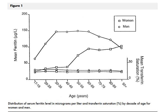
Ferritin, a protein that binds iron, is the most common laboratory measure of iron status. Fertile women have average ferritin levels of around 35 ng/ml, while men in the same age range are much higher, around 150. See the chart above.15xv
At age 45, men have about four times the amount of iron in their bodies as women do, and they also have about four times the rate of heart attacks.
As women reach menopause and cease monthly menstruation, their iron levels rise, and their rates of disease rise also. On average, however, their iron never reaches the levels of men.
The chart shows that average iron levels in men decline starting in the decade of their sixties, and reach a level of around 90 at age 90. This can be attributed to the faster death rate of men with high iron levels and the greater survival of those men with lower iron levels. The amount of iron in each man doesn't change much, only the average in all men.
Men suffer greater rates of cancer, heart disease, and brain-degenerative disorders like Parkinson's and Alzheimer's than women. Iron is implicated in all of these maladies.
It used to be thought that the hormone estrogen in women protected them from heart disease, and that this was the source of the difference in rates of this disease between men and women. But when women undergo a hysterectomy – thus losing the ability to lose iron through the menstrual cycle – heart disease rates increase, and hormone replacement therapy does not affect this.16xvi
If less iron in their bodies explains why women live many years longer than men, as it likely does wholly or partly, that's a very big deal. Iron could represent one of the greatest factors in health, and preventing iron accumulation or lowering high iron stores could be among the most important things anyone could do for their health.
Blood donors are healthier than non-donors
Since blood is the main storage tissue for iron, containing about 80% of total body iron, loss of blood means loss of iron. Blood donors by definition lose blood, so on average they have lower levels of iron than non-donors.
Several scientific studies have looked at the health of blood donors and found huge health benefits to giving blood.
A big issue in studies like these is a selection effect: blood donors on average are likelier to be healthier than non-donors before they ever even donate, since they can't be accepted if they have certain medical conditions, nor would they be as likely to volunteer to donate if they felt unwell. But there are various ways of getting around this limitation, to see whether blood donors are healthier to begin with, whether donating blood makes them healthier, or a combination of both.17
Among a group of nearly 3,000 Finnish men, 153 of them had donated blood at least once in the 24 months preceding the start of the study. The entire group was followed for an average of nine years. In that time, one (0.7%) of the donors had a heart attack, while 316 (12.5%) of the non-donor men had a heart attack. After adjusting for age and all the cardiac risk factors they could think of (cholesterol, weight, etc.), the researchers found that blood donors had an 88% reduced risk of heart attack.xvii
This study tried to adjust for a healthy donor effect by factoring in standard cardiac risk markers, although there might be additional factors that weren't considered.18
In an American population of both men and women who were 40 years old or older and followed for cardiovascular events, those who had donated blood were half as likely to have an event such as a heart attack or stroke.xviii All subjects in this study, both donors and non-donors, had no cardiovascular disease at the start of the study.19
To get around the healthy donor effect, some studies have compared frequent blood donors to infrequent donors. In one such study, those who had given blood at least once in each of three consecutive years were compared to people who only gave blood one time during th20e same period. In the following ten years, frequent donors were only 40% less likely to have a cardiac event as were infrequent donors.xix
Another group of researchers compared blood donors to healthy former blood donors to try to account for the healthy donor effect, and they found that each additional annual blood donation was associated with a 7.5% decreased risk of dying in any given period of time.xx Taken to a logical conclusion, someone who donated blood six times a year – the maximum allowable – would have a 45% lower chance than a non-donor of dying in any given period of time.
This particular study likely greatly underestimates the health benefits of donating blood, since the healthy former donors would on average have lower iron levels than non-donors; therefore, the study compares people currently lowering their iron through blood donation to people who previously lowered their iron stores through blood donation. When compared to people who have never donated blood, the better health of donors would be evident, as we saw above in other studies of blood donation.
The data on blood donors provides compelling evidence that lowering iron lowers health risks. When disease-free donors and disease-free non-donors are followed for the same length of time, the donors are much less likely to become ill or to die, even taking the healthy donor effect into account.
Iron levels are related to total mortality21
The Copenhagen City Heart Study has studied thousands of residents of that city for many years, and is similar to the Framingham Study in the United States. One research group looked at the study's data on almost 9000 people to determine the relation between ferritin (iron) and death rates.xxi
They found that “stepwise increasing concentrations of ferritin were associated with a stepwise increased risk of premature death overall”. People with a ferritin of greater than 600 (a high number) had a median survival age of 55, meaning that of those who had a ferritin that high, half were dead by that age.
Those with a ferritin of 400 to 599 lived an average of 72 years; at 200 to 399, 76 years, and if the ferritin was less than 200, 79 years.
These are startling numbers. Keep in mind that excess iron alone has no symptoms, so many people are walking around with a high ferritin level and don't know it, since only the test shows whether someone has high iron or not.
In the Copenhagen study, 26% of men had ferritin levels greater than 200, while only 6% of women did. The death rate increased from 10 to 15% (depending on cause of death) for every 100 point increase in ferritin.
Although this study didn't calculate it, because of the step-wise increase in mortality, I would expect to find that mortality rates decrease at even lower levels of ferritin, e.g. someone with a ferritin of 100 lives longer than someone with a 200 level, and someone with a level of 50 lives even longer.
Junk iron accumulates as we age
One of the biological hallmarks of aging is the accumulation of cellular “junk”, which causes damage and poor function.
When young, our cells use a regulated process, autophagy, to clear away old and damaged structures and molecules. As we age, this process declines, so that older people have much lower levels of this process. Fasting and certain substances like resveratrol strongly increase autophagy.
Iron creates the toxic waste of aging
In consequence, the cells of older people accumulate22 large amounts of junk. The most important kind of junk is called lipofuscin, which has been termed “the toxic waste of aging”.
Lipofuscin cannot, with some exceptions, be degraded, and its association with age is so strong that scientists consider it a reliable marker of the age of both cells and people. As the amount of lipofuscin grows, it impedes important cell functions, leading to a “garbage catastrophe” - so much accumulated trash that nothing works properly.
Many scientists believe that targeting lipofuscin for clearance is an important way to counteract aging and make cells younger.
Iron is a key player in the formation of lipofuscin.xxii
Iron builds up in cells
Hydrogen peroxide reacts with iron to form lipofuscin
Lipofuscin is almost undegradable
Lipofuscin decreases autophagic capacity
This leads to the accumulation of damaged structures and proteins
Free iron, which is iron that's not locked down by ferritin or other iron storage molecules, is the type that causes damage. The amount of free iron is a function of the amount of total iron, so the less iron in the system altogether, the lower the rate of lipofuscin formation.
Lipofuscin and the iron in it are huge sources of oxidants and can lead to cells becoming senescent, which is a major problem in aging or23ganisms, as the presence of senescent cells causes a system-wi24de increase in oxidative stress, that is, an overabundance of damaging free radicals, which is a prime characteristic of aging.
Senescent cells are loaded with iron, as much as ten times more than younger, better-functioning cells.
When senescent cells are removed from animals using certain drugs, the animals have much better health and live up to 25% longer.
Poorly functioning mitochondria, the cellular structures often called the powerhouses of the cell, can lead to a cell becoming senescent.xxiii Iron accumulation in mitochondria makes them function poorly.xxiv
Decreasing the amount of iron in the body should be effective in preventing the formation of lipofuscin and of senescent cells. If these senescent cells are as important to aging and health as some scientists believe, preventing their formation by keeping iron low could be a huge step in fighting aging.
Growth hormone, fasting, and iron
Scientists believe that one of the ways that calorie restriction slows aging is by reducing the amount of growth hormone, which has been implicated in aging and disease.25
Growth hormone is, as its name implies, a growth factor, and there is a fundamental trade-off between growth and aging. The bigger and faster an organism grows, the faster it ages.
Iron is also a growth factor. Growing animals, including humans, need iron to help make blood, muscle, and other important physiological components.
Growth hormone increases the uptake of iron from food. This makes sense, since when growth hormone signals the body, it wants to grow, and iron is required for growth.
Growth hormone increases iron by decreasing the amount of another hormone, hepcidin, which controls iron uptake from food. When growth hormone was given to human volunteers, it reduced hepcidin concentrations by about two thirds.xxv26
Fasting (going without food for some period of time) retards aging, just as calorie restriction does. Fasting, in contrast to growth hormone, increases the hormone hepcidin, which means that uptake of iron from food decreases.
In short, growth signals cause increased iron uptake and faster aging, and anti-growth signals cause lower iron uptake and slower aging.
So is it growth itself that causes faster aging, or is it the increased iron from growth that does so?
Growth – and growth hormone – also promote aging by activating mTOR, a cellular mechanism crucial to growth, and which also plays a major role in aging. The anti-aging drug rapamycin, which extends life in lab animals, works by deactivating mTOR. However, substances which bind and remove iron (chelators) also deactivate mTOR.xxvi The deactivation of mTOR is prevented by adding iron.
Less iron = lower mTOR activation = longer life and better health.
Exercise lowers iron27
Exercise is known to be one of the most effective health interventions available, and it dramatically l28owers the risk of heart disease, cancer, diabetes, dementia, frailty, and a host of other illnesses.
Many of the health benefits of exercise may be connected to its effect on lowering iron.
Athletes are more likely to suffer from iron deficiency, and this does not seem to be due to diet or excess loss of iron, but to a change in iron uptake.
In the days after a race, marathon runners have a nearly three-fold increase in the iron-regulatory hormone hepcidin. The researchers who found this wrote, “the frequently observed iron deficiency of female runners is caused by elevated hepcidin levels.”xxvii
Lesser amounts of exercise than marathon running also cause elevated levels of hepcidin after exercise.xxviii However, whether low-intensity exercises such as walking affect hepcidin and thus iron does not seem to be known.
As we've seen in this section, many factors that affect aging and health are connected to iron. The fact that they affect iron is probably not the whole story about their health benefits, but is likely very important.
Takeaway Points
In this chapter, we've seen that
calorie restriction, a proven anti-aging intervention, lowers iron levels
preventing iron accumulation in animals makes them live longer
healthful plant compounds, such as quercetin and the substances in green tea, lower iron levels
women live longer than men, and also have lower iron levels
blood donors, who have lower iron levels, are healthier than non-donors
in humans, higher iron (ferritin) associates with a higher death rate
the accumulation of cellular junk is involved in aging, and this junk is loaded with iron
fasting and exercise, which improve health and increase lifespan, lower iron, while growth hormone, which increases aging, also increases iron
References
- Cook, Christopher I., and Byung Pal Yu. "Iron accumulation in aging: modulation by dietary restriction." Mechanisms of Ageing and Development 102.1 (1998): 1-13. ↩
- Reverter-Branchat, Gemma, et al. "Oxidative damage to specific proteins in replicative and chronological-aged Saccharomyces cerevisiae common targets and prevention by calorie restriction." Journal of Biological Chemistry 279.30 (2004): 31983-31989. ↩
- Mori, Nobuko, and Kimiko Hirayama. "Long-term consumption of a methionine-supplemented diet increases iron and lipid peroxide levels in rat liver." The Journal of Nutrition 130.9 (2000): 2349-2355. ↩
- Massie, Harold R., Valerie R. Aiello, and Trevor R. Williams. "Inhibition of iron absorption prolongs the life span of Drosophila." Mechanisms of Ageing and Development 67.3 (1993): 227- 237. ↩
- Schiavi, Alfonso, et al. "Iron-starvation-induced mitophagy mediates lifespan extension upon mitochondrial stress in C. elegans." Current Biology 25.14 (2015): 1810-1822. Klang, Ida M., et al. "Iron promotes protein insolubility and aging in C. elegans." Aging (Albany NY) 6.11 (2014): 975. ↩
- Hatcher, Heather C., et al. "Synthetic and natural iron chelators: therapeutic potential and clinical use." Future Medicinal Chemistry 1.9 (2009): 1643-1670. ↩
- Forte, Giovanni, et al. "Metals in plasma of nonagenarians and centenarians living in a key area of longevity." Experimental Gerontology 60 (2014): 197-206. ↩
- Zacharski, Leo R., et al. "Association of age, sex, and race with body iron stores in adults: analysis of NHANES III data." American Heart Journal 140.1 (2000): 98-104. ↩
- Sullivan, Jerome L. "Are menstruating women protected from heart disease because of, or in spite of, estrogen? Relevance to the iron hypothesis." American Heart Journal 145.2 (2003): 190-194. ↩
- Salonen, Jukka T., et al. "Donation of blood is associated with reduced risk of myocardial infarction The Kuopio Ischaemic Heart Disease Risk Factor Study." American Journal of Epidemiology 148.5 (1998): 445-451. ↩
- Meyers, David G., et al. "Possible association of a reduction in cardiovascular events with blood donation." Heart 78.2 (1997): 188-193. ↩
- Meyers, David G., Kelly C. Jensen, and Jay E. Menitove. "A historical cohort study of the effect of lowering body iron through blood donation on incident cardiac events." Transfusion 42.9 (2002): 1135- 1139. ↩
- Ullum, Henrik, et al. "Blood donation and blood donor mortality after adjustment for a healthy donor effect." Transfusion 55.10 (2015): 2479-2485. ↩
- Ellervik, Christina, et al. "Total and cause-specific mortality by moderately and markedly increased ferritin concentrations: general population study and metaanalysis." Clinical Chemistry 60.11 (2014): 1419-1428. ↩
- Kurz, Tino, Alexei Terman, and Ulf T. Brunk. "Autophagy, ageing and apoptosis: the role of oxidative stress and lysosomal iron." Archives of Biochemistry and Biophysics 462.2 (2007): 220-230. ↩
- Wiley, Christopher D., et al. "Mitochondrial Dysfunction Induces Senescence with a Distinct Secretory Phenotype." Cell Metabolism (2015). ↩
- Walter, Patrick B., et al. "Iron deficiency and iron excess damage mitochondria and mitochondrial DNA in rats." Proceedings of the National Academy of Sciences 99.4 (2002): 2264-2269. ↩
- Troutt, Jason S., et al. "Circulating human hepcidin-25 concentrations display a diurnal rhythm, increase with prolonged fasting, and are reduced by growth hormone administration." Clinical Chemistry 58.8 (2012): 1225-1232. ↩
- Shang, Chaowei, Hongyu Zhou, and Shile Huang. "Iron chelation inhibits mTOR activity in cancer cells." Cancer Research 74.19 Supplement (2014): 2789-2789. ↩
- Roecker, L., et al. "Iron-regulatory protein hepcidin is increased in female athletes after a marathon." European Journal of Applied Physiology 95.5-6 (2005): 569-571. ↩
- Peeling, Peter, et al. "Iron status and the acute post-exercise hepcidin response in athletes." PloS One 9.3 (2014): e93002. ↩
Keep going past iron
Iron is one lever, not the whole machine. P. D. Mangan writes every week about the rest of it: training, protein, insulin, sleep, and the research as it lands.
No charge, and you can unsubscribe at any time. You can also follow P. D. Mangan on X.